Visual neural bridge failed; direct-drive diagnostic passed
Research record
- Question
- Can the official Flyvis visual response drive anatomically labelled left/right descending outputs through an engineered interface into the original full spiking circuit?
- Evidence
- Pinned official visual/spiking sources, v630 and later annotations, and Erginkaya et al. 2025 HS/H2 circuit research. Average broadcasting, later-label transfer and activity-to-rate conversion remain explicit engineering assumptions.
- Experiment
- Linux: 30 fresh full-network trials, 3 fixed seeds and 10 conditions. Two binocular patterns, symmetric motion, grey, input blocks, joint eight-row HS/H2-to-DNp15 cuts and fresh restores. After failure, 9 separate matched direct HS/H2 trials on new seeds; no descending neuron directly stimulated.
- Results
- Both fixed visual-bridge direction criteria fail. DNp15 is right-dominant for A and B on all 3 seeds; DNa02 responds mainly to A. All 6 route cuts give DNp15 0/0 and all 6 restores match whole-network spike events/weights/driver events. Direct HS/H2 A/B criteria pass at DNp15 and DNa02 on all 3 new seeds. Saved-data audits: 352 main checks and 65 diagnostic checks.
- Limitations
- Engineered population means and positive baseline changes discard retinotopy and signed baseline deviations. Conversion is not calibrated biological Hz and differs from Flyvis native rectified synaptic output. Later root-ID label transfer is not independently validated biological identity. Diagnostic input and operating point differ from the visual bridge; the unique failure cause is unresolved. No body, closed-loop autonomy, learning or driving.
- Reproduction
- Frozen plans and local-clock preflight receipts, exact source/checkpoint hashes, both environment locks, all original-network events, main actual driver monitors, diagnostic candidates, raw visual column responses, two auditors, analyses and attempt history in the manifest.
Sources
- Erginkaya et al., Nature Neuroscience 2025
- Original full spiking model at tested commit
- Official visual model at tested commit
- ON and OFF subtype directions, eLife 2018
- Original v630 annotation snapshot
- Later subtype annotation snapshot
Run evidence
Task and result
Connect the tested visual model to the original full spiking circuit without an external steering policy. This first engineered interface failed its fixed direction criterion. Signal reaches the descending outputs; that is not yet reliable direction control.
A separate direct upstream stimulation diagnostic passed. It establishes access to both anatomically labelled output channels at that operating point, not visual perception, autonomous turning or the unique cause of the interface failure.
Interface · assumptions fixed before motor trials
The HS/H2 study used FlyWire snapshot 630, matching our original spiking graph, and identified visual pathways involving HS/H2 and descending cells. Our graph contains all twelve selected HS/H2/DNp15/DNa02 cells. The eight chemical rows from HS/H2 to DNp15 contain 346 model synaptic contacts. These counts come from our pinned model, not a reproduction of the paper’s manually reconstructed matrix. Electrical coupling is not represented.
We generated both moving-edge movies and a grey control with the same official pretrained Flyvis model. All 721 model-column traces of T4a and T4b were saved. Using the source subtype convention, T4a is front-to-back-preferring and T4b back-to-front-preferring; the official visual-model reference directions are 180° and 0° respectively.
Pattern A: left local retinal movie 180°, right 0°. Pattern B: the reverse. The symmetric control uses 180° in both eyes. These are independent local-eye patterns, not a geometrically reconstructed panoramic yaw stimulus.
The projection averages activity across model columns, subtracts the last pre-motion average and keeps positive changes only. It broadcasts each type’s signal to eligible original T4a/b populations. Peak total candidate input is limited to 50,000 Hz per eye, with equalized aggregate scaling across unequal population sizes. The fitted visual weights and native motor-output signs were never tuned using these trial results.
- Identity lookup: 2,701 original-model cells (left a/b: 640/684; right a/b: 662/715). All root IDs occur in the original index and uniquely in the v630 annotation with matching side; 269 later-labelled candidates were excluded. Later type labels are still an annotation-transfer assumption, not independent identity or spatial validation.
- Model assumptions: homogeneous broadcast loses retinotopy. Positive changes in population-mean activity differ from Flyvis’s native per-neuron rectified synaptic output, and discard signed deviations below baseline. The activity-to-pulse conversion is not a measured firing-rate calibration. Native T4 cells use the original author’s refractory-zero stimulation convention; graded biological cells are represented by this model’s spiking equations.
- No external action policy: no goal coordinates, target bearing, route, heading correction, learned driver or fallback steering enters this projection. The original 127,400-neuron / 14,687,178-row spiking graph produces the recorded descending spikes.
Main experiment · 30 fresh full-network trials
Three input-noise seeds × ten conditions: grey, A, B, symmetric motion, A/B input blocks, A/B joint route cuts and A/B fresh restores. Each trial is 1 s at 0.1 ms; motion begins at 200 ms and spikes are counted over 200–1000 ms. The upstream visual samples are 5 ms. Main runner time was 113.62 s on Linux, one CPU thread; the separate visual projection took 6.10 s on the RTX 3060. This is not a speed or model-superiority benchmark.
The criterion was left > right under A and right > left under B on every seed, with DNp15 primary and DNa02 secondary. Neither visual-interface readout passed. No sign flip or output substitution was made.
| Input seed | DNp15 A L / R | DNp15 B L / R | DNa02 A L / R | DNa02 B L / R |
|---|---|---|---|---|
| 20261091 | 1 / 9 | 0 / 30 | 6 / 0 | 0 / 0 |
| 20261092 | 2 / 9 | 0 / 29 | 7 / 1 | 0 / 0 |
| 20261093 | 2 / 9 | 0 / 30 | 8 / 0 | 0 / 0 |
Both input blocks give zero DNp15 and DNa02 spikes. Every joint eight-row route cut gives DNp15 0 / 0. All six fresh restores exactly match the intact whole-network spike-event sequence, weight hash and driver events. Removing this set jointly abolishes the observed DNp15 output in these trials; necessity of each individual edge was not tested. This causal intervention does not establish correct steering.
Direct HS/H2 diagnostic · nine separate trials
After the interface failed, we froze a separate plan and used three new seeds. Four matching 150-Hz candidate channels were mapped to left HS plus opposite H2 for A, and their mirrored targets for B; quiet trials used gate zero. No descending output was directly stimulated. These interventions bypass the earlier visual projection and use a different input population and operating point.
Both DNp15 and DNa02 satisfy the same anatomical side criterion on all three diagnostic seeds; quiet outputs are zero. This demonstrates direct access to both channels under this stimulation, without locating the visual failure uniquely.
| Input seed | DNp15 A L / R | DNp15 B L / R | DNa02 A L / R | DNa02 B L / R |
|---|---|---|---|---|
| 20261101 | 29 / 0 | 0 / 45 | 9 / 8 | 1 / 9 |
| 20261102 | 33 / 0 | 0 / 50 | 9 / 6 | 2 / 7 |
| 20261103 | 32 / 0 | 0 / 50 | 15 / 4 | 0 / 9 |
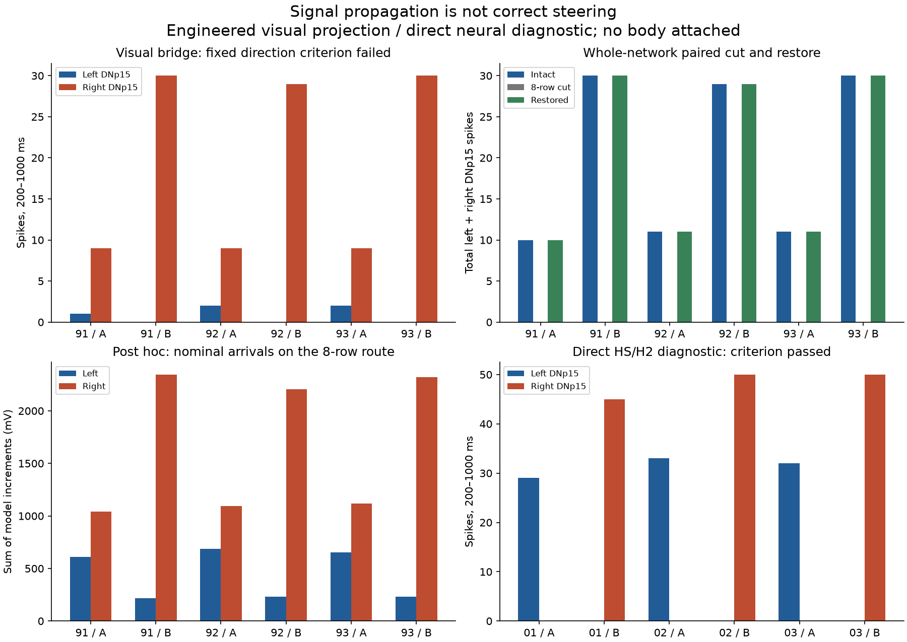
Open the full-size figure. The post hoc arrival panel is model weight × arriving source-spike count, before decay, integration and refractoriness and excluding other inputs. It is not measured current, membrane voltage or a biological causal fraction. The route contains 129 contacts into the left and 217 into the right DNp15; this asymmetry alone has not been isolated as the failure’s cause.
What remains untested
The result supports two narrow statements: visual-model signals can evoke activity through this engineered connection; a separate matched HS/H2 drive can access opposite descending channels. It does not validate the population projection, biological rate conversion, panoramic geometry, physical turning sign, whole-animal behavior or a replacement for the original brain. No closed-loop world, body, learning or driving was attached.
Next, not run: validate native visual-channel specificity and the source’s actual synaptic-output transform before proposing a new projection. Any new test needs a frozen mapping, gain and heldout input seeds. Reversing an output sign, searching gains on these results or attaching a steering policy would not resolve the failure.
Evidence and reproduction
The main NumPy/Pandas auditor passed 352 checks and the diagnostic auditor 65 checks, including event identities/counts, common random schedules, replay/restore criteria and optional live source hashes. A later read-only review repeated 345 main checks and 65 diagnostic checks; the seven optional live Linux source hashes were not repeated in that review. These are saved-data checks, not independent neural reruns or biological validation. Main actual driver monitors are archived; diagnostic candidate schedules are archived, while actual driver equality was asserted inside its runner without separate per-trial monitor files.
Frozen main plan, later diagnostic plan, main summary, diagnostic summary, all output counts, reproduction steps and the complete manifest provide the versions, commands, environment locks and raw evidence. Both models’ learned/original parameters remain unchanged, except for the declared temporary native route cuts and source input convention. All seven original source/data Git blobs match before and after. Public runner copies change only the private baseline path to the equivalent relative location; the hash mapping distinguishes these copies from the unchanged executed originals.
Update record
-
2026-10-28: Executed the engineered visual projection and 30 full-network conditions. Preserved failed direction criteria, input blocks and exact joint-route restorations. Then executed the separately planned nine-trial direct-drive diagnostic with new seeds. Saved-data checks completed; no driving or autonomous steering claim. An initial post hoc table used original weights as references on inactive cut rows; corrected analysis v02 records their actual zero weight. Intact counts, direction criteria and figures’ intact comparisons were unaffected; the earlier analysis remains preserved privately.
-
2026-10-28: Editorial task tracking moved to the native T4 channel follow-up. This preserves the failed visual interface and separate direct-drive diagnostic; it does not imply either was rerun or repaired.